Introduction
Prostate Cancer is the second most diagnosed malignancy in men over the age of ’50′[1]. It is the most common cancer type and is found at autopsy in 30% of men at the age of 50, 40% at age ’60’, and almost 90% at age 90.1 Worldwide, it is the second leading cause of death due to cancer in men, accounting for between 2.1% and 15.2% of all cancer deaths [2]. In Canada, prostate cancer is the third leading cause of cancer death in men; about 10,000 new cases will be diagnosed and about 3,000 men will die from this disease every year [3].
Symptoms due to carcinoma of the prostate are generally absent until extensive local growth or metastases develop, accounting for the fact that only 65% of patients are diagnosed with locally confined disease [2]. Once the tumour has extended beyond the prostate, the risk of metastasis increases dramatically. Tumours smaller than 1 to 1.5 cm3 rarely broach the prostatic capsule [4]. When diagnosed at this early stage, the disease is curable [5]. Even at later stages, treatment can be effective. Nevertheless, treatment options vary depending on the extent of the cancer and prognosis worsens when diagnosis occurs at an advanced stage [4]. Clearly, early diagnosis and accurate staging of prostate cancer, as well as a properly performed therapeutic procedures are critical to the patient’s well being.
Objectives
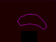
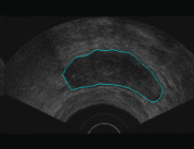
- to learn how to segment images
- to fuse information from different modalities
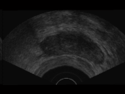
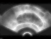
- to investigate MR imaging for prostate
Reference
[1] R.L Waterhouse, M.I Resnick, “The use of transrectal prostatic ultrasonography in the evaluation of patients with prostatic carcinoma.” J. Urol. 141, 233-239, 1989.
[2] E.Silverberg, C. C. Boring, T. S. Squires, “Cancer Statistics, 1990 CA; 40, 9-26, 1990.
[3] Canadian Cancer Statistics 1990, National Cancer Institute of Canada, Toronto, 14, 16, 28, 45, 1990.
[4] M. D. Rifkin, “MRI of the Prostate”. Critical Reviews in Diagnostic Imaging, 31(2), 223-262, 1990.
[5] M.K.Terris, J.E.McNeal, T.A. Stamey, “Estimation of prostate cancer volume by transrectal ultrasound imaging”. J. Urol. 147, 855, 1992.