According to Canadian cancer statistics, in 2004 the most frequently diagnosed cancer in women will continue to be breast cancer. Breast cancer is the second leading cause of death by disease in Canada for women, after lung cancer. In 2004, over 20,000 cases were diagnosed with breast cancer and over 5,200 deaths due to breast cancer were reported, with the death/cases ratio approaching 25%. Early detection of breast cancer, allowing treatment at an earlier stage, can significantly reduce breast cancer mortality.
An x-ray mammogram, ultrasound images and biopsy are the main tools which are utilized for breast cancer diagnosis. MRI-guided biopsy, MR spectroscopy, PET and thermal imaging are also gaining popularity. Recently, many researchers and clinicians are utilizing computer-aided diagnostic (CAD) techniques in order to detect breast cancer. We propose to improve the CAD tools for ultrasound imaging, allowing for more accurate, reliable and rapid detection and diagnosis of breast cancer.
Mammography
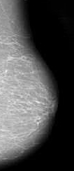
For years, the detection of breast cancer has relied on traditional film (X-ray) mammography. X-ray mammography is currently considered the “gold standard” for breast cancer diagnosis. However, it has been proved ineffective for women with dense breasts. Moreover, it involves radiation, which makes it undesirable. Also, some patients feel discomfort and pain during the procedure. An alternative diagnostic tool to X-ray mammography would be of value.The ultimate diagnosis of all types of breast disease depends on a biopsy. A biopsy is an invasive procedure to remove and examine tissue or cells for the presence of cancer. In most cases the decision for a biopsy is based on mammography findings.
Since biopsy results indicate that 80% of breast lesions detected by mammography are benign, it would be valuable to develop an alternative diagnostic tool, such as CAD ultrasound imaging, for more accurate and rapid detection and diagnosis of breast cancer. This would reduce the number of unnecessary biopsies in patients with benign disease and thus avoid patients’ physical and mental suffering, with an added bonus of reducing healthcare costs.
Ultrasound
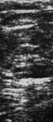
Ultrasound, as an adjunct technique to mammography, can increase the overall sensitivity of conventional breast imaging. There are studies indicating that the use of ultrasound increases cancer detection rate by 14% . Ultrasound imaging of the breast is used to distinguish between solid tumours and fluid-filled cysts. Ultrasound can also be used to evaluate lumps that are hard to see on a mammogram. Recent technological advances have stimulated a revival of interest in the use of ultrasound as a major screening tool, particularly in younger women for whom the risks of radiation from mammography are most significant. Ultrasound imaging is also applicable in a limited fashion to characterize solid masses. However, it is not used as a primary screening tool because of a variable false-negative rate up to 47%, and the operator-dependent nature of efficacy.
Computer-Aided Diagnostic (CAD) Tools
Computer-Aided Diagnostic (CAD) tools involve the use of computers to bring suspicious areas on a mammogram/sonograph to the radiologist’s attention. It is used after the radiologist has done the initial review of the mammogram/sonograph. CAD technology has the potential to improve the accuracy of screening mammography/sonograph.
There is general agreement that ultrasound is an excellent adjunct to mammography. Ultrasound is a safe and acceptable choice for the population being screened. Moreover, it is more cost effective than X-ray mammography. Ultrasound, however, suffers from unacceptably high rates of false negative and false positive results. This shortcoming of ultrasound is mainly due to the difficulty in detecting microcalcifications within speckle noise-corrupted, low-quality images, regardless of the image evaluation being carried out visually by an expert, or automatically by computer algorithms. The low sensitivity and specificity of ultrasound images can certainly be improved with new hardware (higher resolution, colour Doppler) and more efficient/accurate software with intelligent algorithms.
OBJECTIVES
- to enhance/restore the ultrasound images to reduce the effect of speckle noise in order to identify micro-structures more effectively;
- to segment the suspicious structures in the ultrasound image;
- to investigate how to distinguish between malignant and benign disease using 2D target B-mode;
- to learn how to segment images
- to fuse information from different modalities